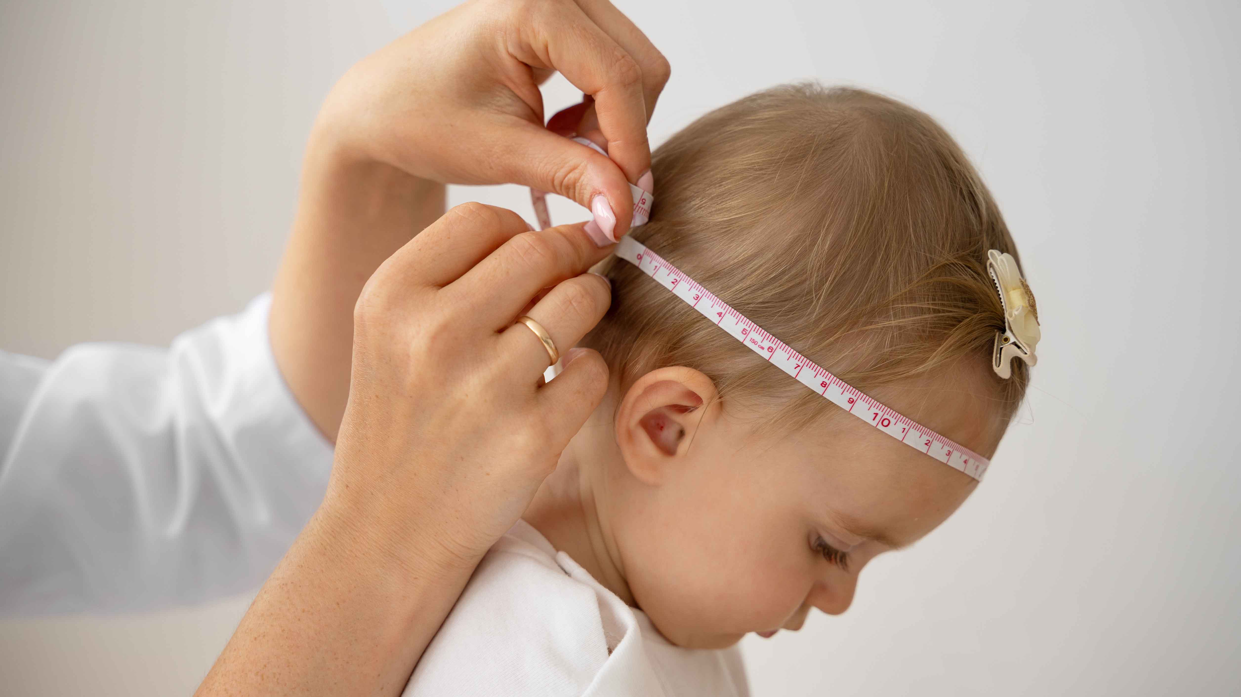
Craniosynostosis is a congenital anomaly that occurs as a result of the early closure of the connective tissues called sutures, which connect the skull bones in infants. Normally, the sutures remain open for a while to allow the baby to grow and the brain to develop. However, in the case of craniosynostosis, one or more of these sutures close prematurely, and a skull deformity occurs. This condition can lead to both aesthetic and functional problems.
In this article written by Neurosurgeon Özgür Taşkapılıoğlu, who serves in Bursa, you can find the causes, types, diagnosis, and treatment methods of craniosynostosis.
What is Craniosynostosis?
Craniosynostosis is the limitation of the growth potential of the skull in certain directions and the formation of an abnormal head shape due to the early closure of sutures. The shape of the skull may vary depending on which suture closes early.
The brain grows rapidly after birth, and this growth requires the movement of the skull bones. Early closure of the sutures can affect brain development and increase intracranial pressure.

What are the Types of Craniosynostosis?
Craniosynostosis is divided into different types according to the closed suture:
1. Sagittal Suture Craniosynostosis (Scaphocephaly)
• It is the most common type.
• Causes elongation of the head in the anterior-posterior direction.
• The head takes on a narrow and long shape.
2. Coronal Suture Craniosynostosis (Plagiocephaly)
• Can be unilateral or bilateral.
• When unilateral, the face and head have an asymmetrical appearance.
• When bilateral, the head takes on a wide and flat shape.
3. Metopic Suture Craniosynostosis (Trigonocephaly)
• Creates narrowing in the forehead area and a triangular head shape.
• May cause the eyes to appear close together.
4. Lambdoid Suture Craniosynostosis
• Rarely seen.
• Creates an asymmetrical deformity in the back of the head.
What are the Causes of Craniosynostosis?
The cause of craniosynostosis is usually unknown, but genetic and environmental factors are thought to play a role.
Genetic Factors:
• Associated with some syndromes (e.g., Apert syndrome, Crouzon syndrome, Pfeiffer syndrome).
• More common in infants with a family history.
Environmental Factors:
• Some drugs or toxins that the mother is exposed to during pregnancy.
• Folic acid deficiency.

What are the Symptoms of Craniosynostosis (Skull Deformity in Infants)?
The symptoms of craniosynostosis may vary depending on which suture closes early:
Abnormal head shape: Flattening or protrusion may occur in certain areas of the head.
Hardening of the skull: While the skull should be soft-textured in infants, a hard area can be felt.
Increased intracranial pressure: In severe cases, restlessness, vomiting, headache, and neurological problems may occur.
Facial asymmetry: Eyes or ears may appear misaligned.
What are the Diagnostic Methods for Craniosynostosis?
Craniosynostosis diagnosis is usually made by physical examination and imaging methods:
1. Physical Examination:
• Examination of skull shape and suture status.
• Evaluation of the baby's developmental characteristics.
• The head takes on a narrow and long shape.
2. Imaging Methods:
• X-ray: Shows early closure of sutures.
• Computed Tomography (CT): Provides three-dimensional imaging of skull bones and brain.
• MR Imaging: Used for more detailed examinations.
3. Genetic Tests:
• Genetic tests can be performed in cases of suspected syndromic craniosynostosis.
What are the Treatment Methods for Craniosynostosis?
The treatment of craniosynostosis is usually surgical. The treatment plan depends on the severity of the condition, the child's age, and general health.
1. Surgical Intervention
• Cranial Reconstruction: Opening of sutures and reshaping of the skull.
• Endoscopic Surgery: A less invasive method and is usually applied in early diagnosed cases.
2. Skull Shaping Helmets
• Can be used after surgery or in mild cases to guide the natural growth of the skull.
Importance of Early Diagnosis and Intervention in Craniosynostosis
Early diagnosis is vital in the treatment of craniosynostosis. Especially in infants whose brain development continues, early initiation of treatment positively affects long-term results.
What Happens if Craniosynostosis is Not Treated?
• Increased intracranial pressure.
• Developmental delay.
• Permanent skull deformities.
Recommendations for Families for Craniosynostosis
Infant Follow-up: Observe your baby's head shape and general development from birth.
Expert Opinion: Consult a pediatric neurosurgeon when you notice an abnormal condition.
Post-Surgical Rehabilitation: Do not neglect the controls and practices recommended by the doctor after surgery.
Craniosynostosis is a condition that can be successfully managed with early diagnosis and appropriate treatment. It is of great importance for families to make careful observations and seek expert help so that children can maintain their physical and mental development in a healthy way. If you notice a skull deformity in your baby, you should consult a specialist without delay. Prof. Dr. Özgür Taşkapılıoğlu wishes you healthy days.
Make an Appointment Now
+90 530 167 07 40